
pestis with the assistance of the Caf1M chaperon ( 18, 19). In the course of its biogenesis, F1 polymers are generated in the periplasm of Y. The expression of the caf1 operon is optimal at 37☌, i.e., upon the translocation of the pathogen from the flea vector to the mammalian host ( 17). An important component of these anti-plague subunit vaccines is the Caf1 (F1) capsule antigen that is expressed from the caf1 operon on the pMT-1 103Kb plasmid of Y. Although several vaccine candidates confer protection in animal models of plague and in clinical studies, none of them has been approved for use in Western countries ( 14). pestis strains resistant to multiple therapeutic antibiotic ( 12, 13) and the fear of a natural or intentional disease outbreak initiated by antibiotic-resistant strains emphasize the need to develop vaccines against this deadly disease. Plague morbidity and mortality rates have substantially decreased since the introduction of antimicrobials, but the isolation of Y. pestis is classified as a potential bioterror agent ( 11).

Because of its lethality and infectivity, Y. Plague is an infectious disease caused by the Gram-negative bacterium Yersinia pestis, which has claimed the lives of millions of people throughout human history ( 10). These data open avenues for urgently needed effective antibacterial vaccines.
Kozak sequence full#
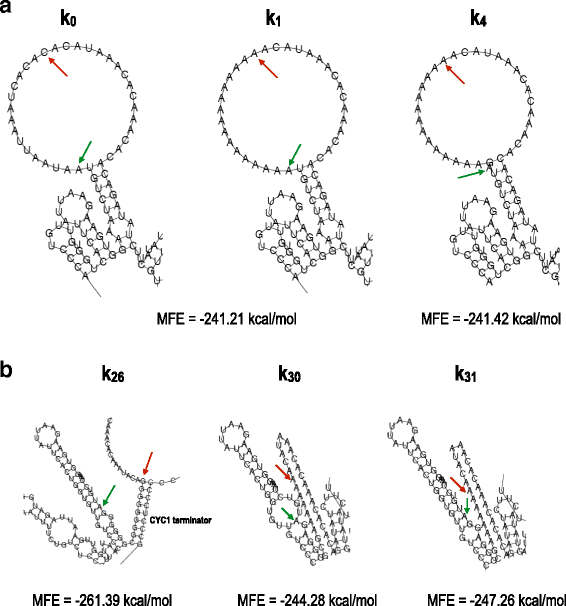
Our mRNA-LNP vaccine elicited humoral and cellular immunological responses in C57BL/6 mice and conferred rapid, full protection against lethal Y. Now, the disease is treated effectively with antibiotics however, in the case of a multiple-antibiotic-resistant strain outbreak, alternative countermeasures are required. Plague is a rapidly deteriorating contagious disease that has killed millions of people during the history of humankind. We designed a nucleoside-modified mRNA-LNP vaccine based on the bacterial F1 capsule antigen, a major protective component of Yersinia pestis, the etiological agent of plague. Here, we developed an effective mRNA-LNP vaccine against a lethal bacterial pathogen by optimizing mRNA payload guanine and cytosine content and antigen design. Although currently applied toward viral pathogens, data concerning the platform’s effectiveness against bacterial pathogens are limited. For initiation of translation from such a site, other features are required in the mRNA sequence in order for the ribosome to recognize the initiation codon.Messenger RNA (mRNA) lipid nanoparticle (LNP) vaccines have emerged as an effective vaccination strategy. Lmx1b is an example of a gene with a weak Kozak consensus sequence. There are examples in vivo of each of these types of Kozak consensus, and they probably evolved as yet another mechanism of gene regulation. The cc at -1 and -2 are not as conserved, but contribute to the overall strength. An 'adequate' consensus has only 1 of these sites, while a 'weak' consensus has neither. either A or G in the consensus) relative to the number 1 nucleotide must both match the consensus (there is no number 0 position). For a 'strong' consensus, the nucleotides at positions +4 (i.e. The A nucleotide of the "AUG" is referred to as number 1. Some nucleotides in this sequence are more important than others: the AUG is essential since it is the actual initiation codon encoding a methionine amino acid at the N-terminus of the protein. In vivo, this site is often not matched exactly on different mRNAs and the amount of protein synthesized from a given mRNA is dependent on the strength of the Kozak sequence. The Kozak sequence is not to be confused with the ribosomal binding site (RBS), that being either the 5' cap of a messenger RNA or an Internal Ribosome Entry Site (IRES). The ribosome requires this sequence, or a possible variation (see below) to initiate translation. This sequence on an mRNA molecule is recognized by the ribosome as the translational start site, from which point a protein is coded by that mRNA molecule. Better weighing performance in 6 easy steps


 0 kommentar(er)
